

Abstract
As part of the therapeutic evaluation, positron emission tomography/computed tomography (PET/CT) using 18F-fluorodeoxyglucose (18F-FDG) was performed on a 39-year-old patient with metastatic melanoma of the left thigh who was subsequently receiving immunotherapy. The examination revealed pathologic hypermetabolic foci in the lymph nodes and liver along with a highly suspicious pathologic hypermetabolic foci in the spinal marrow at the level of the first lumbar vertebra (L1). The presence of such a hypermetabolic focus can significantly decrease survival duration, highlighting the importance of early detection. PET/CT with 18F-FDG proved to be more sensitive and specific than CT alone in identifying occult distant metastases, as the latter may underestimate malignant involvement of the spinal marrow.
Keywords: PET-18F-FDG, melanoma, spinal marrow, metastasis
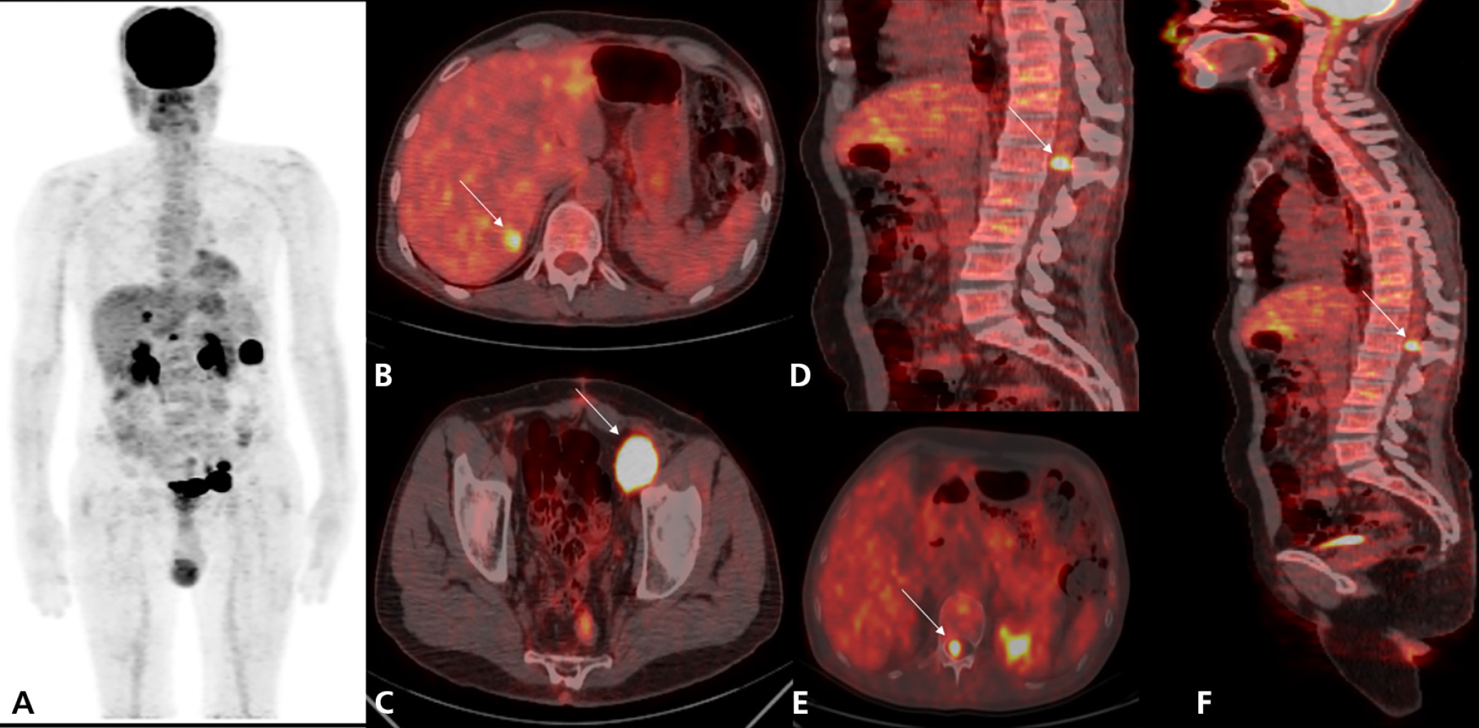
Malignant melanoma is considered one of the most lethal cancers (1), with a poor prognosis once metastasized. A rare site of metastasis for melanoma is the heart, spleen, and spinal marrow, which can significantly reduce survival time, underscoring the importance of early detection (2). Follow-up examinations using 18F-FDG PET/CT have been employed to assess intramedullary lesions, especially in tumors characterized by high-grade malignancy, such as melanoma (3), and ideally must be diagnosed early and confirmed through dedicated magnetic resonance imaging (2).
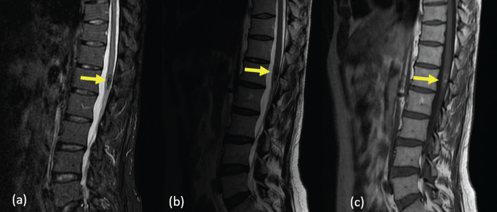
The disease course was characterized by the rapid onset of neurological symptoms a few months after diagnosis, followed by worsening and eventual demise.
Regular imaging follow-up is essential for monitoring therapeutic effectiveness and detecting new metastases in patients with malignant melanoma (4). However, there is significant variability in surveillance methods due to limited scientific data. Although no randomized trials have compared follow-up approaches with or without PET/CT imaging, a meta-analysis indicated PET/CT was superior to CT alone in identifying distant metastases, with higher sensitivity (86% vs. 63%) and specificity (91% vs. 78%) (2).
After conducting a comprehensive literature review, we found no similar cases of medullary metastasis from malignant melanoma demonstrated by PET/CT. To the best of our knowledge, we are the first case of this type. Our case highlights the role of 18F-FDG PET/CT in the follow-up of metastatic melanoma by early diagnosis of spinal metastases, even before the onset of clinical symptoms. This underscores the importance of conducting further studies to compare the various imaging modalities used in the therapeutic assessment of metastatic malignant melanoma, particularly at atypical sites, as well as to evaluate their contributions.
Footnotes
Ethics
Informed Consent: Informed consents of the patient was obtained.
Authorship Contributions
Concept: C.B., S.O.N., K.B., O.A.S., Y.B., A.D., Design: C.B., S.O.N., O.A.S., Y.B., A.D., Data Collection or Processing: C.B., Analysis or Interpretation: C.B., S.O.N., A.D., Literature Search: C.B., Writing: C.B.
Conflict of Interest: No conflicts of interest were declared by the authors.
Financial Disclosure: The authors declare that this study has received no financial support.
References
Tsao H, Atkins MB, Sober AJ. Management of cutaneous melanoma. N Engl J Med. 2004;351:998-1012.
Vercellino L, Rivas A, Baroudjian B, Lebbé C, Merlet P. Role of FDG PET in the evaluation of locoregional and distant spread of melanoma staging. Nuclear Medicine. 2020;44:305-312.
Merhemic Z, Stosic-Opincal T, Thurnher MM. Neuroimaging of spinal tumors. Magn Reson Imaging Clin N Am. 2016;24:563-579.
Bier G, Hoffmann V, Kloth C, Othman AE, Eigentler T, Garbe C, La Fougère C, Pfannenberg C, Nikolaou K, Klumpp B. CT imaging of bone and bone marrow infiltration in malignant melanoma--challenges and limitations for clinical staging in comparison to 18FDG-PET/CT. Eur J Radiol. 2016;85:732-738.

