
Multiple Bone Involvement in Low-grade Myofibroblastic Sarcoma Demonstrated on 18F-FDG PET/CT
PDF

Interesting Image

Multiple Bone Involvement in Low-grade Myofibroblastic Sarcoma Demonstrated on 18F-FDG PET/CT

Mol Imaging Radionucl Ther 2024;33(3):206-208 DOI: 10.4274/mirt.galenos.2024.14238
, , , ,
PDF
Permissions

Abstract

A 68-year-old woman with low back pain for 2 months was admitted. T2-weighted fat-saturated imaging revealed hyperintense lesions in multiple lumbar regions, suggesting the possibility of bone metastases. Multiple osteolytic and mixed osteolytic-osteoblastic lesions with significant 18F-fluorodeoxyglucose (18F-FDG) uptake, as well as multiple osteoblastic lesions with mild 18F-FDG uptake, were observed on subsequent 18F-FDG positron emission tomography/computed tomography without an identifiable primary lesion. This patient was pathologically diagnosed with low-grade myofibroblastic sarcoma (LGMS) after biopsy and surgery. Although multiple bone involvement in LGMS is extremely rare, this case suggests that it should be considered in the differential diagnosis of multiple bone metastases.

Keywords: Low-grade myofibroblastic sarcoma, bone destruction, 18F-FDG PET/CT
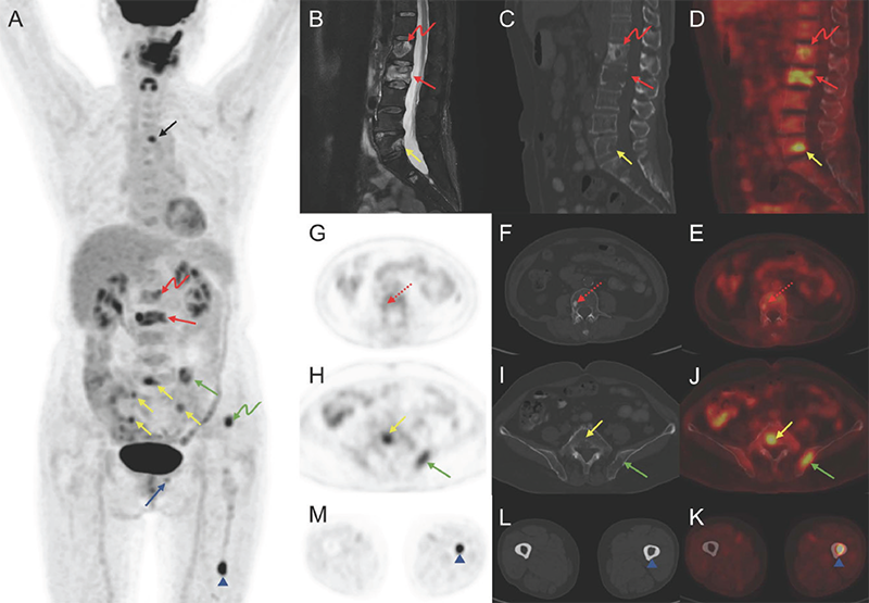
Figure 1
Figure 1. A 68-years-old woman with low back pain for 2 months was admitted. T2-weighted fat-saturated imaging revealed heterogeneous hyperintense lesions in multiple lumbar regions, indicating the likelihood of bone metastases. To identify the primary lesion and determine the tumor stage, the 18F-fluorodeoxyglucose (18F-FDG) positron emission tomography/computed tomography (PET/CT) was performed (A, MIP; B, sagittal T2-weighted fat-saturated magnetic resonance imaging; C, sagittal CT; D, sagittal PET/CT; E, J, K, axial PET/CT; F, I, L, axial CT; G, H, M, axial PET). Multiple osteolytic (solid arrow) and mixed osteolytic-osteoblastic (bend arrow) lesions with significant 18F-FDG uptake, as well as multiple osteoblastic lesions (dotted arrow) with mild 18F-FDG uptake, were observed in the thoracic vertebra (black arrow), lumbar vertebra (red arrow), sacrum (yellow arrow), right 12th rib, left ilium (green arrow), left pubis (blue arrow), and femur (arrowhead), without an identifiable primary lesion. In addition, intense activity was found in the oral cavity and left cheek area, with no abnormalities detected on the corresponding CT scans, indicating unspecific uptake. This patient underwent biopsy of the left iliac region and surgery of L3 and was pathologically diagnosed with low-grade myofibroblastic sarcoma (LGMS). LGMS is a rare mesenchymal tumor that was previously thought to mainly originate from the head and neck region, but a recent study suggested it may be more common in the extremities (1,2). The majority of LGMS occurs in the soft tissue and is infrequent in the bone (2,3). The distal femur is the most prevalent site for bone LGMS, followed by the ilium, and the vertebrae are rare (3,4,5). LGMS has been described as having a low-grade malignant potential that may locally recur and less frequently metastases (2). Multifocal involvement is extremely rare, with only one case of multicentric soft tissue involvement described by Wechalekar et al. (6). We report an extremely rare LGMS case of multiple bone involvement (multiple vertebrae, rib, ilium, pubis, and femur) without any soft tissue lesions. Furthermore, in this case, different types of bone destruction (osteolytic, mixed osteolytic-osteoblastic, and osteoblastic) were observed, which is quite different from the primary and metastatic bone lesions in the previous studies, which typically manifest as osteolytic bone destruction (1,4,5,7,8). 18F-FDG PET/CT scan is an important approach in differentiating malignant sarcomatous lesions from benign lesions, especially for the equivocal lesions on conventional imaging, as well as a primary diagnostic tool for metastatic lesion detection (9,10). The presence of primary bone LGMS on the 18F-FDG PET/CT scan has been reported by Hou et al. (8). To the best of our knowledge, this is the first case of LGMS involving numerous bones and causing different types of bone destruction with heterogeneous 18F-FDG uptake on the 18F-FDG PET/CT. Despite its rarity, this case suggests that LGMS with multiple bone involvement should be considered when multiple bone metastases are suspected.
View larger Download

Footnotes

Ethics

Informed Consent: The written informed consent has been obtained from the patient.

Authorship Contributions

Concept: L.S., W.Z., Design: L.S., W.Z., Data Collection or Processing: X.H., Analysis or Interpretation: X.H., Literature Search: H.L., N.G., Writing: H.L.

Conflict of Interest: No conflicts of interest were declared by the authors.

Financial Disclosure: This work was supported by the Key Clinical Projects of Peking University Third Hospital (no.: BYSY2022060).

References

1

San Miguel P, Fernández G, Ortiz-Rey JA, Larrauri P. Low-grade myofibroblastic sarcoma of the distal phalanx. J Hand Surg Am. 2004;29:1160-1163.

2

Chan JY, Gooi Z, Wong EW, Ng SK, Tong MC, Vlantis AC. Low-grade myofibroblastic sarcoma: A population-based study. Laryngoscope. 2017;127:116-121.

3

Hadjigeorgiou GF, Samaras V, Varsos V. Low-grade myofibroblastic sarcoma of the thoracic spine: report of an extreme rare case. Br J Neurosurg. 2017;31:731-733.

4

Watanabe K, Ogura G, Tajino T, Hoshi N, Suzuki T. Myofibrosarcoma of the bone: a clinicopathologic study. Am J Surg Pathol. 2001;25:1501-1507.

5

Saito T, Mitomi H, Kurisaki A, Torigoe T, Takagi T, Suehara Y, Okubo T, Kaneko K, Yao T. Low-grade myofibroblastic sarcoma of the distal femur. Int J Surg Case Rep. 2013;4:195-199.

6

Wechalekar MD, Ayres O, Farshid G, Clayer M, Cleland LG. Multicentric myofibroblastic sarcoma. BMJ Case Rep. 2014;2014:bcr2013201666.

7

Wang L, Li LX, Chen DQ, Yang L, Li SK, Cheng C. Low-grade Myofibroblastic sarcoma: clinical and imaging findings. BMC Med Imaging. 2019;19:36.

8

Hou W, Su M, Li Q, Tian R. Low-Grade Myofibroblastic Sarcoma Demonstrated on 99mTc-MDP Bone Scan and 18F-FDG PET/CT. Clin Nucl. Med. 2020;45:549-551.

9

Mendoza H, Nosov A, Pandit-Taskar N. Molecular imaging of sarcomas with FDG PET. Skeletal Radiol. 2023;52:461-475.

10

Roberts CC, Kransdorf MJ, Beaman FD, Adler RS, Amini B, Appel M, Bernard SA, Fries IB, Germano IM, Greenspan BS, Holly LT, Kubicky CD, Lo SS, Mosher TJ, Sloan AE, Tuite MJ, Walker EA, Ward RJ, Wessell DE, Weissman BN. ACR Appropriateness Criteria Follow-Up of Malignant or Aggressive Musculoskeletal Tumors. J Am Coll Radiol. 2016;13:389-400.

