

Abstract
Paragangliomas (PGLs) are neuroendocrine tumors originating from the neural crest. They usually arise from the adrenal medulla and sympathetic or parasympathetic ganglions. Approximately 10% of PGLs are located in the extra-adrenal gland. Renal PGL is a rare condition. In this case report, we present the 18F-fluorodeoxyglucose positron emission tomography/computed tomography (PET/CT) and 68Ga-DOTATATE PET/CT findings of polycystic kidney-derived PGL.
Keywords: Renal paraganglioma, 18F-FDG PET/CT, 68Ga-DOTATATE PET/CT
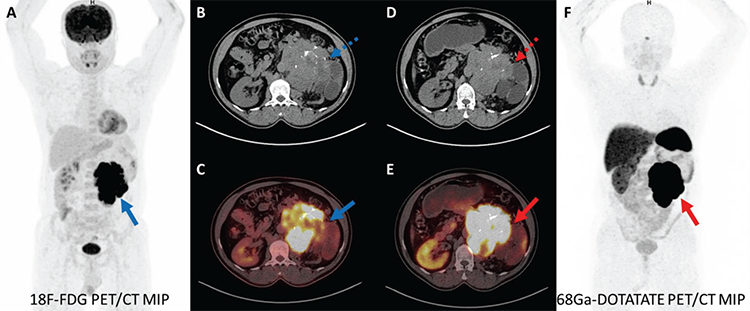
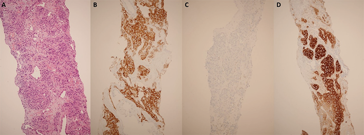
Extra-adrenal PGLs most commonly occur in the carotid body, vagal body, mediastinum, and retroperitoneum (1). Renal PGL is rare (1,2). In the genitourinary tract, the renal pelvis (4.9%) is the third primary site of PGLs, followed by the bladder (79.2%) and urethra (12.7%) (3). The clinical symptoms depend on the location of the mass in the kidney (2,4). Functional imaging methods play an important role in confirming diagnosis, staging, and restaging (5). PGLs contain high levels of somatostatin receptors (6,7). In a study evaluating 22 PGL patients with 68Ga-DOTATATE PET/CT, the detection rate of lesions was 100% (8). In another study evaluating 23 patients with PGLs using 18F-FDG PET/CT and 68Ga-DOTATATE PET/CT, many regions were 68Ga-DOTATATE and 18F-FDG positive, but 18F-FDG uptake was lower than 68Ga-DOTATATE uptake (median SUVmax values were 12.5-21, respectively) (9). 68Ga-DOTATATE PET/CT has higher diagnostic accuracy than 18F-FDG PET/CT (10). Renal PGL should be considered when evaluating kidney masses because it can mimic RCC, and caution should be exercised.
Footnotes
Ethics
Informed Consent: The patient consent was obtained.
Authorship Contributions
Surgical and Medical Practices: Z.T., S.B.K., N.E., T.F.Ç., E.A., Concept: Z.T., S.B.K., T.F.Ç., E.A., N.E., Design: Z.T., N.E., S.B.K., T.F.Ç., E.A., Data Collection or Processing: Z.T., S.B.K., N.E., T.F.Ç., E.A., Analysis or Interpretation: Z.T., S.B.K., N.E., T.F.Ç., E.A., Literature Search: Z.T., S.B.K., N.E., T.F.Ç., E.A., Writing: Z.T., T.F.Ç., E.A., S.B.K., N.E.
Conflict of Interest: No conflicts of interest were declared by the authors.
Financial Disclosure: The authors declare that this study has received no financial support.
References
Neumann HPH, Young WF Jr, Eng C. Pheochromocytoma and Paraganglioma. N Engl J Med. 2019;381:552-565.
Bahar B, Pambuccian SE, Gupta GN, Barkan GA. Renal paraganglioma: report of a case managed by robotic assisted laparoscopic partial nephrectomy and review of the literature. Case Rep Urol. 2014;2014:527592.
Peng C, Bu S, Xiong S, Wang K, Li H. Non-functioning paraganglioma occurring in the urinary bladder: A case report and review of the literature. Oncol Lett. 2015;10:321-324.
Yi C, Han L, Yang R, Yu J. Paraganglioma of the renal pelvis: a case report and review of literature. Tumori. 2017;103(Suppl 1):e47-e49.
Taïeb D, Timmers HJ, Hindié E, Guillet BA, Neumann HP, Walz MK, Opocher G, de Herder WW, Boedeker CC, de Krijger RR, Chiti A, Al-Nahhas A, Pacak K, Rubello D; European Association of Nuclear Medicine. EANM 2012 guidelines for radionuclide imaging of phaeochromocytoma and paraganglioma. Eur J Nucl Med Mol Imaging. 2012;39:1977-1995.
Sobocki BK, Perdyan A, Szot O, Rutkowski J. Management of Pheochromocytomas and Paragangliomas: A Case-Based Review of Clinical Aspects and Perspectives. J Clin Med. 2022;11:2591.
Şahin R, Baykal Koca S, Yücetaş U, Çermik TF, Ergül N. 18F-FDG PET/CT and 68Ga-DOTATATE PET/CT Findings in a Patient With Primary Renal Well-Differentiated Neuroendocrine Tumor. Clin Nucl Med. 2022;47:e503-e505.
Skoura E, Priftakis D, Novruzov F, Caplin ME, Gnanasegaran G, Navalkissoor S, Bomanji J. The impact of Ga-68 DOTATATE PET/CT imaging on management of patients with paragangliomas. Nucl Med Commun. 2020;41:169-174.
Chang CA, Pattison DA, Tothill RW, Kong G, Akhurst TJ, Hicks RJ, Hofman MS. (68)Ga-DOTATATE and (18)F-FDG PET/CT in Paraganglioma and Pheochromocytoma: utility, patterns and heterogeneity. Cancer Imaging. 2016;16:22.
Carrasquillo JA, Chen CC, Jha A, Ling A, Lin FI, Pryma DA, Pacak K. Imaging of Pheochromocytoma and Paraganglioma. J Nucl Med. 2021;62:1033-1042.

