

Abstract
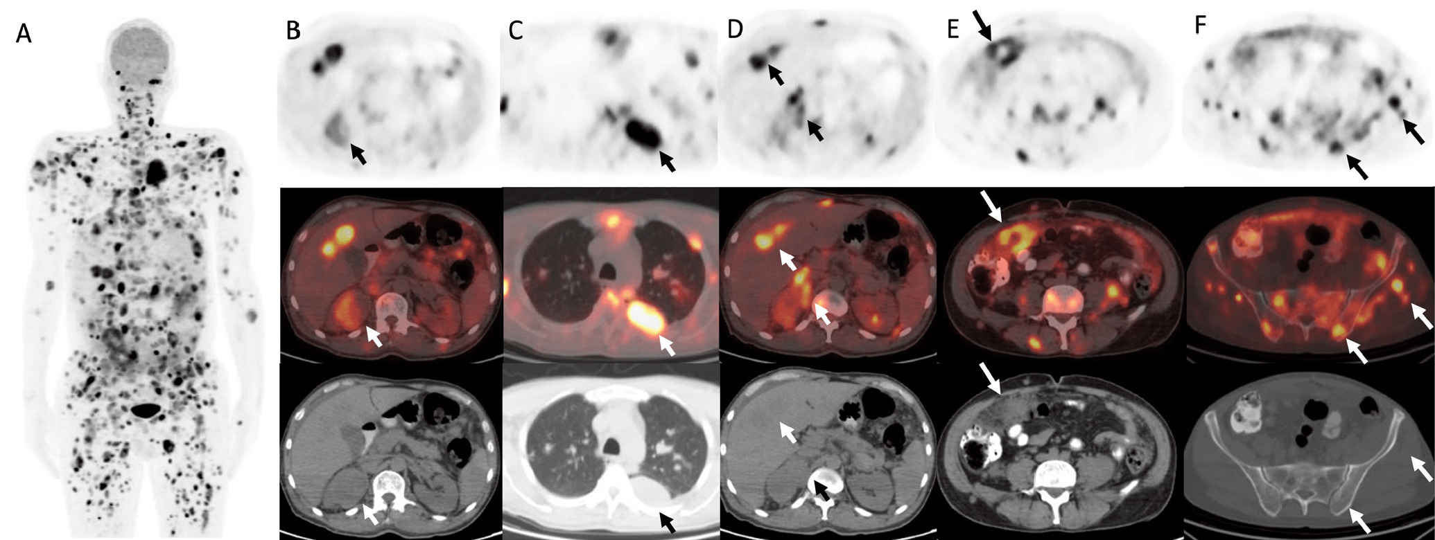
The diagnostic performance of 18F-fluorodeoxyglucose (18F-FDG) positron emission tomography/computed tomography (PET/CT) for primary kidney tumors is limited. Nevertheless, 18F-FDG PET/CT is valuable for staging renal cell carcinoma (RCC) when suspected metastases coexist, as one-third of patients with RCC have distant metastases upon diagnosis. Herein, we present a 53-year-old male patient with extensive 18F-FDG-avid metastatic lesions and an 18F-FDG-avid renal mass, which later revealed RCC.
Keywords: 18F-FDG PET/CT, papillary renal cell carcinoma, metastasis, staging
Footnotes
Ethics
Informed Consent: Patient consent was obtained.
Authorship Contributions
Surgical and Medical Practices: M.O., M.Y.K., Concept: M.O., D.H.Ş., Y.Ş., Design: M.O., D.H.Ş., Y.Ş., Data Collection or Processing: M.O., M.Y.K., Analysis or Interpretation: M.O., S.K., Literature Search: M.O., S.K., Writing: M.O.
Conflict of Interest: No conflicts of interest were declared by the authors.
Financial Disclosure: The authors declare that this study has received no financial support.
References
Liu Y. The Place of FDG PET/CT in Renal cell carcinoma: value and limitations. Front Oncol. 2016;6:201.
Karivedu V, Jain AL, Eluvathingal TJ, Sidana A. Role of positron emission tomography imaging in metabolically active renal cell carcinoma. Curr Urol Rep. 2019;20:56.
Wang HY, Ding HJ, Chen JH, Chao CH, Lu YY, Lin WY, Kao CH. Meta-analysis of the diagnostic performance of [18F]FDG-PET and PET/CT in renal cell carcinoma. Cancer Imaging. 2012;12:464-474.
Mendhiratta N, Muraki P, Sisk AE Jr, Shuch B. Papillary renal cell carcinoma: review. Urol Oncol. 2021;39:327-337.
Connor Wells J, Donskov F, Fraccon AP, Pasini F, Bjarnason GA, Beuselinck B, Knox JJ, Rha SY, Agarwal N, Bowman IA, Lee JL, Pal SK, Srinivas S, Scott Ernst D, Vaishampayan UN, Wood LA, Simpson R, De Velasco G, Choueiri TK, Heng DYC. Characterizing the outcomes of metastatic papillary renal cell carcinoma. Cancer Med. 2017;6:902-909.
Xuan D, Wen W, Tian S, Piao M, Xu D, Liu L. Prognostic value of maximum standard uptake value, metabolic tumor volume, and total lesion glycolysis of 18F-FDG PET/CT in patients with renal carcinoma: a protocol for systematic review and meta analysis. Medicine (Baltimore). 2020;99:e19988.
Pankowska V, Malkowski B, Wedrowski M, Wedrowska E, Roszkowski K. FDG PET/CT as a survival prognostic factor in patients with advanced renal cell carcinoma. Clin Exp Med. 2019;19:143-148.
Wu C, Cui Y, Zhao Y, Chen X, Liao X, Di L, Yin L, Liu M, Wang R. Elevated tumor-to-liver standardized uptake value ratio (TLR) from preoperative 18F-FDG PET/CT predicts poor prognosis of patients with clear cell renal cell carcinoma after nephrectomy. Eur J Radiol. 2020;131:109218.

