

Abstract
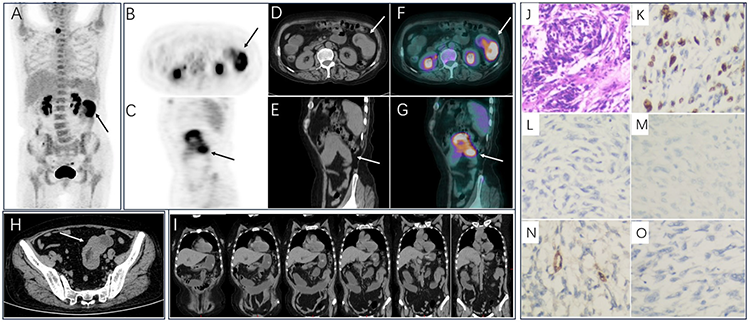
Spindle cell sarcoma is a malignant tumor with low incidence. They can occur in the soft tissue, bone, or viscera. The characteristics of morphology, density, and metabolism of spindle cell sarcoma are related to the location of the lesion. A 61-year-old woman presented with vomiting after eating for 2 weeks. Signs of peritoneal irritation were involved, but no response for symptomatic treatment included antiemetic and antispasmodic therapy. Abdominal computed tomography (CT) indicated a mass in the intestinal tract in the pelvic cavity. Then, 18F-fluorodeoxyglucose (18F-FDG) positron emission tomography/CT was performed, which interestingly detected a jejunal malignancy mass in the left upper abdomen with annular high uptake of 18F-FDG, which was complicated by intussusception and intestinal obstruction. Finally, the jejunal mass was pathologically clarified as an undifferentiated spindle cell sarcoma.
Keywords: 18F-FDG PET/CT, jejunum, undifferentiated spindle cell sarcoma, intussusception, intestinal obstruction
Ethics
Informed Consent: We obtained informed consent from study subjects for publishing their data.
Authorship Contributions
Concept: X.L., Design: X.L., Data Collection or Processing: H.L., Analysis or Interpretation: H.L., Literature Search: X.L., Writing: H.L.
Conflict of Interest: No conflicts of interest were declared by the authors.
Financial Disclosure: This work was supported by foundation of “Lvyang Jinfeng” talents attracting plan (LYJF00040) and project of Jiangsu provincial health commission (H2023055) and Jiangsu shuangchuang talent as well as the Jiangsu provincial medical key discipline cultivation unit (JSDW202251).
Footnotes
Ethics
Informed Consent: We obtained informed consent from study subjects for publishing their data.
Authorship Contributions
Concept: X.L., Design: X.L., Data Collection or Processing: H.L., Analysis or Interpretation: H.L., Literature Search: X.L., Writing: H.L.
Conflict of Interest: No conflicts of interest were declared by the authors.
Financial Disclosure: This work was supported by foundation of “Lvyang Jinfeng” talents attracting plan (LYJF00040) and project of Jiangsu provincial health commission (H2023055) and Jiangsu shuangchuang talent as well as the Jiangsu provincial medical key discipline cultivation unit (JSDW202251).
References
Fisher C. Spindle cell sarcomas. Surg Pathol Clin 2011;4:721-744.
Bouvier AM, Robaszkiewicz M, Jooste V, Cariou M, Drouillard A, Bouvier V, Nousbaum JB; French Network of Cancer Registries (FRANCIM). Trends in incidence of small bowel cancer according to histology: a population-based study. J Gastroenterol 2020;55:181-188.
Lambrinow J, Świątkowski F, Jurga M, Gajdzis P, Chabowski M. Intussusception caused by an extremely rare tumor: undifferentiated spindle cell sarcoma. Pol Arch Intern Med 2023;133:16491.
Cheng WH, Chen JJ, Tsou YA, Tseng GC. Jejunal undifferentiated spindle cell sarcoma with glossal metastasis. Kaohsiung J Med Sci 2012;28:120-121.
Gao YH, Huang JJ, Chen P. A case of non-gastrointestinal stromal spindle cell sarcoma of the small intestine. Chin J Gastrointest Surg 2018;21:1181-1182.
Hong SM, Jung SH, Baek DH. Diagnostic yields and clinical impacts of capsule endoscopy. Diagnostics (Basel) 2021;11:1842.
Treglia G, Bongiovanni M, Paone G, Ceriani L, Giovanella L. Metastatic undifferentiated spindle cell sarcoma of the thyroid gland evaluated by 18F-FDG PET/CT. Clin Nucl Med 2015;40:e208-e210.
Wu X, Zhang C, Huang Y, Wang H, Jiang L. Maxillofacial spindle cell sarcoma with lung metastases on FDG PET/CT imaging. Clin Nucl Med 2018;43:703-704.

