
Late 68Ga PSMA-positive Pancreatic Metastasis From Renal Cell Carcinoma in a Patient with Metastatic Prostate Cancer: A Mission Impossible
PDF

Interesting Image

Late 68Ga PSMA-positive Pancreatic Metastasis From Renal Cell Carcinoma in a Patient with Metastatic Prostate Cancer: A Mission Impossible

Mol Imaging Radionucl Ther 2024;33(2):112-114 DOI: 10.4274/mirt.galenos.2023.90582
, , , ,
PDF
Permissions

Abstract

We present the case of a patient with newly diagnosed high-risk prostate cancer. The patient underwent nephrectomy for renal cell carcinoma (RCC) in 2009. The prostate-specific membrane antigen (PSMA) scan revealed a primary tumor with seminal vessel involvement, PSMA-positive regional lymph nodes, several nodular lung lesions with mild PSMA uptake, PSMA-positive mediastinal lymph nodes, and a PSMA-positive mass in the pancreatic head. Ultrasound-guided biopsy was performed for the pancreatic lesions revealing metastasis from a RCC. Simultaneous treatment for prostate cancer and metastatic RCC was initiated. To separate metastatic sites for both primaries, we attempted to use fluorodeoxyglucose positron emission tomography/computed tomography, which was moderately positive for the pancreatic mass but not for the other locations. RCC is a 68Ga PSMA-positive tumor; the synchronous combination of RCC with prostate cancer can be confusing and requires more complex clinical interpretation.

Keywords: PSMA, FDG, PET/CT, prostate cancer, RCC, pancreatic metastasis

Ethics

Informed Consent: The patient has consented to the use of his medical record for scientific purposes.

Authorship Contributions

Surgical and Medical Practices: A.D., P.B., Concept: P.B., Design: P.B., Data Collection or Processing: G.M., N.N., P.B., Analysis or Interpretation: G.M., N.N., M.G.-T., P.B., Literature Search: P.B., Writing: G.M., P.B.

Conflict of Interest: No conflicts of interest were declared by the authors.

Financial Disclosure: The authors declare that this study has received no financial support.
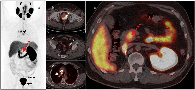
Figure 1
Figure 1. 68Ga prostate-specific membrane antigen (PSMA) scan performed for staging of prostate cancer [Gleason score of 7 (4+3), grade group 3, initial prostate-specific antigen (PSA) 206 ng/mL] [a, maximum intensity projection (MIP) image], revealing a highly positive primary prostate tumor (b, arrow-axial hybrid image) with parailiac lymph node metastasis (c, dashed arrow-axial hybrid image), a PSMA-positive paratracheal lymph node, and a round, well-shaped lung metastasis (d, open arrow-axial hybrid image). PSMA-positive lesion in the pancreatic head (e, red arrow-axial hybrid image). The patient underwent nephrectomy for renal cell carcinoma (RCC) in 2009 and was considered to be in remission. Knowing that the pancreas is a possible location for late RCC metastasis and can be PSMA-positive (1,2), we recommended an ultrasound-guided biopsy for the pancreatic lesion, which proved infiltration by a typical clear cell renal carcinoma, PAX 8 and GATA3 positive, although RCC negative on immunohistochemistry. Simultaneous treatment for prostate cancer and metastatic RCC was initiated as follows: hormone therapy maximal androgen blockade for prostate cancer and bevacizumab/interferon, later changed to pazopanib for RCC.
View larger Download
Figure 2
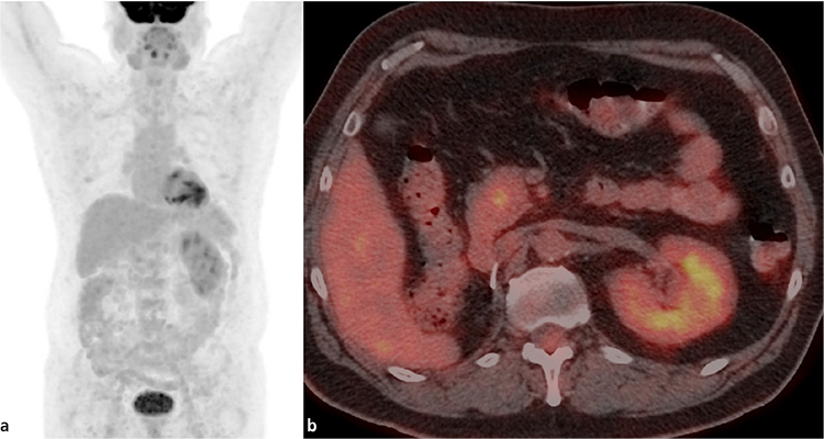
Figure 2. Fluorodeoxyglucose (FDG) positron emission tomography scan: pancreatic lesion is faintly FDG positive. The other lesions did not show substantial FDG uptake (should be borne on mind that the patient is under treatment at the time of the FDG scan). a. 18F-FDG MIP image; b. 18F-FDG axial hybrid imaging faint FDG uptake in the pancreatic lesion. To separate metastatic sites for both primaries, we attempted to use FDG PET/computed tomography (CT) (Figure 2) as a discriminator, as proposed in the literature (3). The scan was obtained after starting treatment for both and at PSA levels of 3.24 ng/mL. FDG was moderately positive for the pancreatic mass but not for the other locations (lung nodules were small volume) (Figure 2). On a size basis, there was no change in the pancreatic mass, but there was a reduction in the lung, mediastinal, and primary prostate sites, which additionally confused the interpretation. However, as a final decision, the patient had partial response for prostate cancer and non-progression (partial response or stable disease) for RCC and remained on the same treatment. This actually shows the inability of separating metastatic locations for all imaging modalities used (68Ga PSMA, FDG, and CT) and the need for an individual approach and more complex clinical interpretation of the case.
View larger Download

Footnotes

Ethics

Informed Consent: The patient has consented to the use of his medical record for scientific purposes.

Authorship Contributions

Surgical and Medical Practices: A.D., P.B., Concept: P.B., Design: P.B., Data Collection or Processing: G.M., N.N., P.B., Analysis or Interpretation: G.M., N.N., M.G.-T., P.B., Literature Search: P.B., Writing: G.M., P.B.

Conflict of Interest: No conflicts of interest were declared by the authors.

Financial Disclosure: The authors declare that this study has received no financial support.
References

1

Katsourakis A, Noussios G, Hadjis I, Alatsakis M, Chatzitheoklitos E. Late solitary pancreatic metastasis from renal cell carcinoma: a case report. Case Rep Med 2012;2012:464808.

2

Demirci E, Ocak M, Kabasakal L, Decristoforo C, Talat Z, Halaç M, Kanmaz B. (68)Ga-PSMA PET/CT imaging of metastatic clear cell renal cell carcinoma. Eur J Nucl Med Mol Imaging 2014;41:1461-1462.

3

Tariq A, Kwok M, Pearce A, Rhee H, Kyle S, Marsh P, Raveenthiran S, Wong D, McBean R, Westera J, Dunglison N, Esler R, Navaratnam A, Yaxley JW, Thomas P, Pattison DA, Roberts MJ. The role of dual tracer PSMA and FDG PET/CT in renal cell carcinoma (RCC) compared to conventional imaging: A multi-institutional case series with intra-individual comparison. Urol Oncol 2022;40:66.e1-66.e9.

