

Abstract
Nowadays, the utility of positron emission tomography/computed tomography (PET/CT) is well established in nasopharyngeal carcinoma (NPC). The incidence of NPC in the West population, especially in children, is very low. We present the first Italian case of a pediatric patient with NPC followed up with 18F-fluorodeoxyglucose (18F-FDG) PET/CT scan in addition to the standard follow-up imaging methods, including CT and magnetic resonance imaging. The 18F-FDG PET/CT scan was helpful in discriminating between metastatic and benign osseous lesions, thereby helping clinicians to determine the most appropriate therapeutic regimen. These findings support the clinical utility of 18F-FDG PET/CT in the diagnostic work-up of pediatric patients with NPC.
Keywords: Positron emission tomography, 18F-fluorodeoxyglucose, nasopharyngeal carcinoma, pediatrics
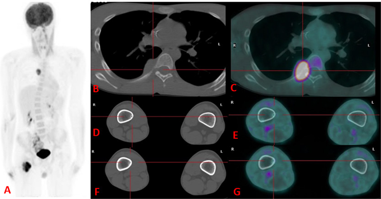
An additional 18F-FDG PET/CT was conducted, highlighting increased uptake in the intertrochanteric and lesser trochanter lesions, whereas the other two distal diaphysis lesions showed no significant 18F-FDG uptake. Furthermore, a further hypermetabolic lesion (SUVmax: 8.4) was noted in the right-sided sixth costovertebral joint. On the left box (A) maximum intensity projection image showing 18F-FDG-avid metastases in the intertrochanteric and lesser trochanter regions of the right femur and in the sixth right-sided rib, with corresponding CT and fuzed PET/CT images in boxes B and C, respectively. An additional 18F-FDG-avid lesion involving the great and lesser trochanter of the right femur was observed, whereas tiny lesions seen on CT images were confirmed to be enchondromas with no significant FDG uptake (D, E, F, G).
Based on the PET/CT findings, chemotherapy with gemcitabine and oxaliplatin was administered, and radiation therapy (30 Gy) was delivered to the proximal region of the right femur. After 5 months, post-therapy PET/CT revealed the disappearance of the hypermetabolic lesions in keeping with a complete metabolic response to therapy. Since then, the boy has been followed up by two further 18F-FDG PET/CT scans with no evidence of disease recurrence. The lesions in the distal diaphysis of the right femur were followed up by MRI and after 1 year remained unchanged, in keeping with enchondromas, according to the radiologist.
The incidence of NPC in the Western countries is low (especially in the pediatric age) compared with that in South-Eastern Asia (one vs. 8 per 100,000 person per year) (,). Although most nasopharyngeal lesions in the pediatric population are benign, the possibility of NPC should be considered when symptoms last long (). Current imaging methods used to assess NPC at diagnosis and follow-up include CT, MRI, and bone scan. To the best of our knowledge, this is the first pediatric case of NPC evaluated by 18F-FDG PET/CT reported in Italy. 18F-FDG PET/CT emerged as an additional tool because of its metabolic information, helping characterize doubtful findings at conventional imaging (). In our case, 18F-FDG PET/CT played a crucial role in characterizing the enchondromas in the distal femur, as these well-defined lesions on the MRI scan demonstrated no significant uptake of 18F-FDG, consistent with previous findings in the literature. Only three pediatric case reports (,,) and one original study () including exclusively pediatric patients with NPC have been published in western countries so far. In that original study, Cheuk et al. () proved in a group of 86 children that MRI is superior to 18F-FDG PET/CT in the local staging and detection of local lymph node metastases. Nevertheless, 18F-FDG PET/CT was useful to address the correct nature of ambiguous findings suspected for distant metastases and to estimate complete disease remission at 3-6 months after months, a timeline earlier than that achievable with MRI ().
Footnotes
Ethics
Informed Consent: Informed consent was obtained from all patients included in the study.
Authorship Contributions
Surgical and Medical Practices: S.I., S.P., A.P., Concept: D.M.M., Design: S.P., Data Collection or Processing: V.L.L., A.M.M., Analysis or Interpretation: S.I., A.P., G.M.G., Literature Search: N.Q., Writing: N.Q., D.M.M.
Conflict of Interest: No conflicts of interest were declared by the authors.
Financial Disclosure: The authors declare that this study has received no financial support.
References
Gatta G. Epidemiological Aspects in Nasopharyngeal Cancer. In: Vermorken JB, Budach V, Leemans CR, Machiels J-P, Nicolai P, O’Sullivan B (eds.) Critical Issues in Head and Neck Oncology. Cham, Springer 2021;319-325.
Boussen H, Bouaouina N, Mokni-Baizig N, Gamoudi A, Chouchane L, Benna F, Ladgham A. Les carcinomes du nasopharynx: donnée actuelles [Nasopharyngeal carcinoma. Recent data]. Pathol Biol (Paris) 2005;53:45-51.
Ben-Ami T, Kontny U, Surun A, Brecht IB, Almaraz RL, Dragomir M, Pourtsidis A, Casanova M, Fresneau B, Bisogno G, Schneider DT, Reguerre Y, Bien E, Stachowicz-Stencel T, Österlundh G, Wygoda M, Janssens GO, Zsiros J, Jehanno N, Brisse HJ, Gandola L, Christiansen H, Claude L, Ferrari A, Rodriguez-Galindo C, Orbach D. Nasopharyngeal carcinoma in children and adolescents: The EXPeRT/PARTNER diagnostic and therapeutic recommendations. Pediatr Blood Cancer 2021;68:e29018.
Cheuk DK, Sabin ND, Hossain M, Wozniak A, Naik M, Rodriguez-Galindo C, Krasin MJ, Shulkin BL. PET/CT for staging and follow-up of pediatric nasopharyngeal carcinoma. Eur J Nucl Med Mol Imaging 2012;39:1097-1106.
Stolten M, Moak S, Chauhan A, Warrier R. Facial pain in a child with attention deficit hyperactivity disorder. Clin Pediatr (Phila) 2016;55:196-198.
Muzaffar R, Vacca F, Guo H, Mhapsekar R, Osman MM. Pediatric nasopharyngeal carcinoma as seen on 18F-FDG PET/CT. Front Oncol 2019;9:110.
Stokken J, Manz RM, Flagg A, Kate Gowans L, Anne S. Synchronous occurrence of nasopharyngeal carcinoma and Hodgkin lymphoma. Int J Pediatr Otorhinolaryngol 2014;78:154-156.

