

Abstract
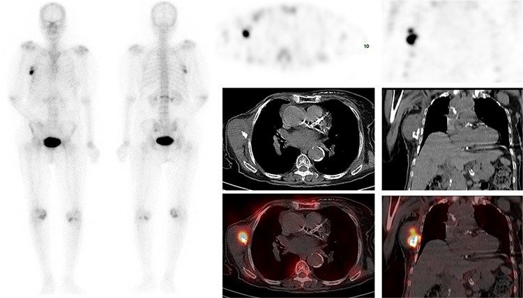
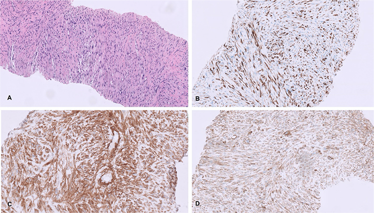
Tc-99m methylene diphosphonate (MDP) is a bone imaging agent used for skeletal staging, but it can also be localized in extraosseous calcifying lesions. We report a case of an 84-year-old woman with breast carcinoma who underwent surgery followed by radiotherapy 10 years ago and now presented with a right axillary mass referred for Tc-99m MDP to exclude bone metastasis. Tc-99m MDP shows intense tracer uptake in the right thoracic region corresponding to the site of calcified soft tissue mass in the right lateral chest wall. Subsequent ultrasonography revealed an ill-defined lesion containing coarse calcifications. Biopsy showed radiation-induced sarcoma. Extra osseous Tc-99m MDP uptake may provide important diagnostic information that may alter patient management.
Keywords: Tc-99m MDP bone scan, extraosseous uptake, radiation-induced sarcoma, breast cancer
Radiation-induced sarcoma (RIS) is a rare type of cancer that develops in the radiation field of a previous radiation therapy treatment. It is a long-term complication of radiation therapy that typically occurs years after the initial treatment (). The exact mechanisms underlying the development of RISs are not fully understood. However, radiation exposure can cause genetic mutations or damage to DNA within normal cells, leading to the development of cancerous cells over time. The incidence rates of RISs in breast cancer patients typically range from 0.03% to 0.3% (3 to 30 cases per 10,000 patients) over a period of 10 to 20 years following radiation therapy (). One study reported a cumulative incidence of 0.07% 5 years after irradiation, 0.27% at 10 years, and 0.48% at 15 years; the 5-year survival rate was 36% ().
Tc-99m MDP is a bone imaging agent used for skeletal staging, but it can also be localized in extraosseous calcifying lesions. The reported mechanisms include (a) local tissue necrosis or damage leading to increased calcium deposition in the tissue, (b) hyperemia, (c) altered capillary permeability, (d) adsorption onto tissue calcium, (e) presence of iron deposits, and (f) binding to enzyme receptors or denatured proteins (, ). Significantly increased MDP uptake by extraosseous soft tissue tumors is associated with poor differentiation in both children and adults (). Extra-osseous tracer uptake is common on Tc-99m MDP, and these non-osseous findings may occasionally alter diagnosis and subsequent management. In hybrid imaging, anatomical localization with computed tomography often improves both the specificity of assessment of the bone lesion and soft tissue uptake. Clinical history and familiarity with typical imaging appearances are crucial for distinguishing soft tissue masses.
Ethics
Informed Consent: Informed consent was obtained from the patient.
Authorship Contributions
Surgical and Medical Practices: S.U., A.J., K.A.R., Z.N., S.K., V.V.J., Concept: S.U., A.J., K.A.R., Design: S.U., Data Collection or Processing: S.U., S.K., V.V.J, Analysis or Interpretation: S.U., A.J., Z.N., Literature Search: S.U., Writing: S.U., A.J., K.A.R.
Conflict of Interest: No conflicts of interest were declared by the authors.
Financial Disclosure: The authors declare that this study has received no financial support.
Footnotes
Ethics
Informed Consent: Informed consent was obtained from the patient.
Authorship Contributions
Surgical and Medical Practices: S.U., A.J., K.A.R., Z.N., S.K., V.V.J., Concept: S.U., A.J., K.A.R., Design: S.U., Data Collection or Processing: S.U., S.K., V.V.J, Analysis or Interpretation: S.U., A.J., Z.N., Literature Search: S.U., Writing: S.U., A.J., K.A.R.
Conflict of Interest: No conflicts of interest were declared by the authors.
Financial Disclosure: The authors declare that this study has received no financial support.
References
Hall EJ, Wuu CS. Radiation-induced second cancers: the impact of 3D-CRT and IMRT. Int J Radiat Oncol Biol Phys 2003;56:83-88.
Di Lalla V, Tolba M, Khosrow-Khavar F, Baig A, Freeman C, Panet-Raymond V. Radiation-induced sarcomas of the breast: a review of a 20-year single-center experience. Cureus 2023;15:e38096.
Kirova YM, Vilcoq JR, Asselain B, Sastre-Garau X, Fourquet A. Radiation-induced sarcomas after radiotherapy for breast carcinoma: a large-scale single-institution review. Cancer 2005;104:856-863.
Usmani S, Ahmed N, Muzaffar S, Al Kandari F. Extensive extramedullary hematopoiesis in thalassemia: soft tissue uptake on 99mTc-MDP SPECT/CT. Clin Nucl Med 2020;45:e459-e460.
Usmani S, Gnanasegaran G, Marafi F, Esmail A, Ahmed N, Van den Wyngaert T. The clinical significance of incidental soft tissue uptake on whole body 18F-sodium fluoride bone PET-CT. Clin Radiol 2019;74:95-110.
Liu S, Xie J, Yu F, Cai H, Wu F, Zheng H, Ma C, Lv Z, Wang H. 99mTc-methylene diphosphonate uptake in soft tissue tumors on bone scintigraphy differs between pediatric and adult patients and is correlated with tumor differentiation. Cancer Manag Res 2020;12:2449-2457.

