
Misdiagnosis of a Drain-site Hernia Containing Fallopian Tube Fimbria on 18F-FDG PET/CT
PDF

Interesting Image

Misdiagnosis of a Drain-site Hernia Containing Fallopian Tube Fimbria on 18F-FDG PET/CT

Mol Imaging Radionucl Ther 2024;33(2):115-117 DOI: 10.4274/mirt.galenos.2023.17894
, , , ,
PDF
Permissions

Abstract

In a 55-year-old woman with sigmoid colon cancer, a subcutaneous mass in the left lower abdomen was incidentally found and gradually enlarged. For further diagnosis and staging, an 18F-fluorodeoxyglucose (FDG) positron emission tomography/computed tomography scan was performed, which revealed a subcutaneous mass in the left lower abdomen with mild uptake of 18F-FDG, suggesting the possibility of metastasis. However, post-surgery and pathological confirmation, this mass was diagnosed as a drain-site hernia containing fallopian tube fimbria, which is extremely rare but should be considered in the differential diagnosis of subcutaneous mass in the lower abdomen.

Keywords: Subcutaneous mass, 18F-FDG PET/CT, drain-site hernia, fallopian tube fimbria

Ethics

Informed Consent: Written informed consent has been obtained from the patient.

Authorship Contributions

Surgical and Medical Practices: B.Q.L., Concept: W.Z., Design: W.Z., Data Collection or Processing: N.G., Analysis or Interpretation: L.S., Literature Search: H.L., L.S., Writing: H.L.

Conflict of Interest: No conflict of interest was declared by the authors.

Financial Disclosure: This work was supported by the Key Clinical Projects of Peking University Third Hospital (no: BYSY2022060).
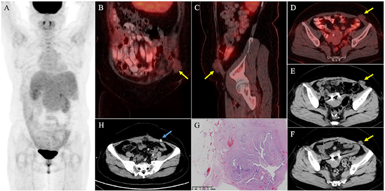
Figure 1
Figure 1. A 55-year-old woman with sigmoid colon cancer underwent laparoscopic sigmoid colectomy three years ago. Eight months after colectomy, she underwent right hepatectomy and chemotherapy because of newly diagnosed liver metastasis. Eight months after chemotherapy, the patient incidentally found a subcutaneous palpable hard mass in the left lower abdomen with slight intermittent abdominal discomfort. During follow-up, the mass gradually enlarged, while serum carcinoembryonic antigen levels remained within the normal range (2.6 to 3.3 ng/mL, normal reference range: 0-5.0 ng/mL). Twelve months after the discovery of the mass, an 18F-fluorodeoxyglucose (FDG) positron emission tomography/computed tomography (PET/CT) scan was conducted for further diagnosis and staging [(A) maximum intensity projection, (B) coronal PET/CT, (C) sagittal PET/CT, (D) axial PET/CT], revealing an irregularly shaped subcutaneous mass in the left lower abdomen (B, C, and D, yellow arrow). The mass was closely related to the abdominal wall (without a remarkable defect) and showed mild uptake of 18F-FDG (maximum standardized uptake value: 1.2). We then reviewed the serial CT scans (E and F, 6 and 12 months before the PET/CT scan), which demonstrated that the mass enlarged and became irregular. Therefore, the possibility of metastasis was raised. However, post-surgery and pathological confirmation (G, fallopian tube tissue could be seen in the fibrous and fatty tissue), this mass was finally diagnosed as a drain-site hernia (DSH) containing fallopian tube fimbria. We reviewed all previous CT scans of the patient and confirmed that the mass was indeed located above the former 5 mm drain site (H, blue arrow). In addition, the mass appeared six months post-colectomy and gradually enlarged, but it was missed by the radiologist. This misdiagnosis should serve as a reminder of the following three lessons. First, we should enhance and broaden our understanding of rare hernias. Trocar site hernia (TSH) is rare and is defined as an incisional hernia occurring at the trocar incision site after laparoscopic surgery (1). Among TSH, DSH is an even rarer type, occurring at the port site where the drainage tube is placed (2). Its prevalence ranges from 0.1% to 3.4% according to the literature. Trocar size is the dominant risk factor for DSH, which often occurs at the ≥10 mm port site and rarely occurs at the 5 mm port site (as observed in this patient) (3,4,5). Second, we need to broaden and improve our knowledge of the atypical contents of hernias. The most common contents of hernias are the small bowel and omentum. The uncommon hernia contents, such as the fallopian tube in this case, appendix, ovary, gall bladder, and bladder, are rare but cannot be ignored (2,6,7,8). It should be noted that any organ within the abdominal cavity might herniate. DSH-containing fallopian tubes are extremely rare, with only three cases reported (6,7,8). Finally, a thorough review of previous clinical image data and patient history is essential for accurate diagnosis. If we could comprehensively review all previous CT scans, this misdiagnosis might be avoided. In summary, although a DSH containing the fallopian tube fimbria is extremely rare, this misdiagnosed case suggests that it should be considered in the differential diagnosis of a subcutaneous mass in the lower abdomen.
View larger Download

Footnotes

Ethics

Informed Consent: Written informed consent has been obtained from the patient.

Authorship Contributions

Surgical and Medical Practices: B.Q.L., Concept: W.Z., Design: W.Z., Data Collection or Processing: N.G., Analysis or Interpretation: L.S., Literature Search: H.L., L.S., Writing: H.L.

Conflict of Interest: No conflict of interest was declared by the authors.

Financial Disclosure: This work was supported by the Key Clinical Projects of Peking University Third Hospital (no: BYSY2022060).

References

1

Gao X, Chen Q, Wang C, Yu YY, Yang L, Zhou ZG. Rare case of drain-site hernia after laparoscopic surgery and a novel strategy of prevention: A case report. World J Clin Cases 2020;8:6504-6510.

2

Makama JG, Ameh EA, Garba ES. Drain Site Hernia: A Review of the Incidence and Prevalence. West Afr J Med 2015;34:62-68.

3

Su J, Deng C, Yin HM. Drain-site hernia after laparoscopic rectal resection: A case report and review of literature. World J Clin Cases 2022;10:2637-2643.

4

Nacef K, Chaouch MA, Chaouch A, Khalifa MB, Ghannouchi M, Boudokhane M. Trocar site post incisional hernia: about 19 cases. Pan Afr Med J 2018;29:183.

5

Helgstrand F, Rosenberg J, Bisgaard T. Trocar site hernia after laparoscopic surgery: a qualitative systematic review. Hernia 2011;15:113-121.

6

Sharma L, Singh A, Bhaskaran S, Radhika AG, Radhakrishnan G. Fallopian tube herniation: an unusual complication of surgical drain. Case Rep Obstet Gynecol 2012;2012:194350.

7

Ergin A, Işcan Y, Ağca B, Karip B, Memisoğlu K. Fallopian tube herniation from trocar-site after laparoscopic appendectomy. Ulus Travma Acil Cerrahi Derg 2020;26:639-641.

8

Hussain K, Masood J. Fallopian Tube Herniation through Left Sided Abdominal Drain Site. J Coll Physicians Surg Pak 2016;26:S19-20.

