

Abstract
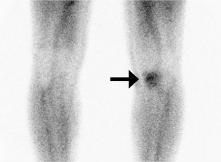
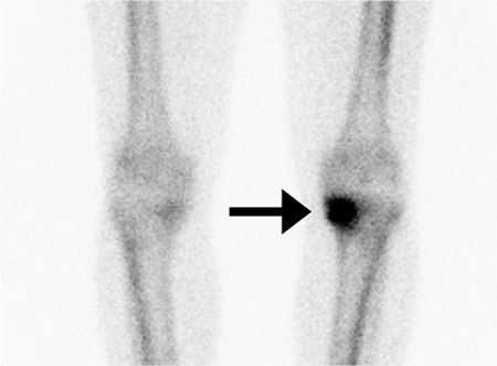
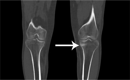
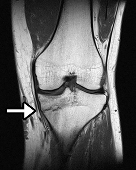
Stress fractures are injuries most commonly seen in the lower limbs and are usually caused by repetitive stress. While the distal and middle third of the tibia is the most frequent site for stress fractures (almost 50%), stress fractures of the proximal tibia is relatively rare and could be confused with other types of tibial fractures, thus altering management plans for the clinician. Early diagnosis of stress fractures is also important to avoid complications. Imaging plays an important role in the diagnosis of stress fractures, especially bone scan. Combined with single-photon emission computed tomography/computed tomography (SPECT/CT) it is an important imaging technique for stress fractures in both upper and lower extremities, and is widely preferred over other imaging techniques. In this case, we present the case of a 39-year-old male patient diagnosed with stress fracture of the proximal tibia and demonstrate the contribution of CT scan fused with SPECT imaging in the early diagnosis of stress fracture prior to other imaging modalities.
Keywords: Stress fracture, proximal tibia, bone scintigraphy, single-photon emission computed tomography/computed tomography
Footnotes
Ethics
Informed Consent: Consent form was filled out by all participants.
Peer-review: Externally peer-reviewed.
Authorship Contributions
Surgical and Medical Practices: B.O., N.C., Concept: B.O., N.C., Design: B.O., N.C., Data Collection or Processing: B.O., N.C., Analysis or Interpretation: B.O., N.C., Literature Search: P.A., Writing: N.C.
Conflict of Interest: No conflict of interest was declared by the authors.
Financial Disclosure: The authors declared that this study received no financial support.

