

Abstract
Osteopoikilosis is an inherited condition with autosomal dominant trait resulting in sclerotic foci throughout the skeleton. It has been suggested that loss-of-function mutations of LEMD3 gene located on 12q14.3 result in osetopoikilosis. A bp heterozygote deletion was detected in our patient at the cytosine nucleotide at position 1105 with molecular genetic analysis. Although this mutation has not been previously described, it was considered to be the most likely cause of the disease in our patient due to frame shift and premature stop codon formation. As in our case, three phase bone scintigraphy and whole body imaging did not reflect the true extent of lesion sites and lesion activity. SPECT/CT images could reflect lesion location and activity more accurately, and could be a good alternative for differential diagnosis of unexplained bone pain and sclerotic lesions in one examination.
Keywords: LEMD3 geni, osteopoikiloz, Tc-99m MDP, SPECT/BT
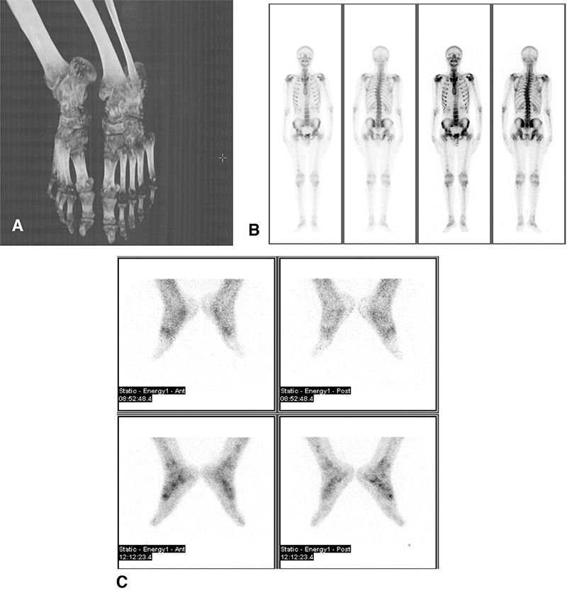
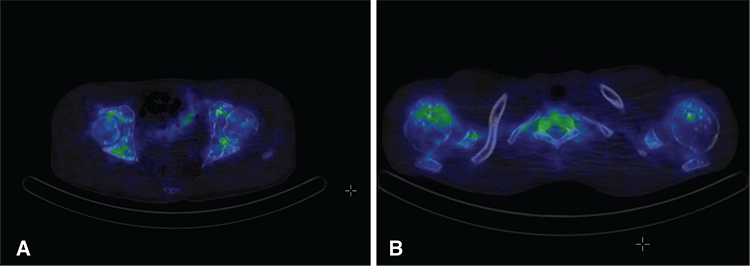
Osteopoikilosis is an inherited condition with autosomal dominant trait that results in numerous sclerotic foci throughout the skeleton (1). Previous studies have reported that these lesions are symmetric but randomly distributed, with increased concentrations at the carpal and tarsal bones, as well as at the ends of the long bones (2,3). Lesions are reported to be located less in the skull, rib, vertebra, and mandible (1).
Recently, whole-genome linkage analysis of affected individuals resulted in identification of a loss-of-function mutation in gene LEMD3 at position 12q14.3. LEMD3 is believed to function in bone morphogenetic protein (BMP) signaling by interacting with the family of SMAD proteins downstream from transforming growth factor-beta (TGF-beta) to regulate bone formation (4,5). It has been reported that LEMD3 can antagonize both BMP and TGF-beta signaling in human cells (6). Molecular genetic analysis was performed in our patient, and a bp heterozygote deletion was detected in the cytosine nucleotide at position 1105. This mutation has not been previously described in the literature, but is considered to be the most likely cause of the disease due to frame shift and premature stop codon formation.
Histologically, the sclerotic foci correspond to old and inactive remodeling of spongiform lamellar trabeculae with a gross nodular or star-like appearance (7). Sclerotic lesions are classically inactive on whole body scintigraphy (8,9). For this reason, a Tc-99m bone scan is sufficiently diagnostic and usually returns to normal in cases of osteopoikilosis. There have been a few cases reported in the literature of abnormal bone scans in patients with osteopoikilosis (10). In such cases, the pattern of increased uptake is usually symmetric and localized to the distal ends of tubular, carpal, and tarsal bones. This may correspond to active bone remodeling of these multiple foci, which has also been reported in pathologic specimens of osteopoikilosis. As in our patient, localized three phase bone scintigraphy and whole body scanning did not reflect the true extent of lesion site and activity. However, SPECT/CT images could reflect lesion location and activity more accurately, and could be a good alternative for differential diagnosis of unexplained bone pain and sclerotic lesions in one examination.
Footnotes
Ethics
Informed Consent: Consent form was filled out by all participants.
Peer-review: Externally peer-reviewed.
Authorship Contributions
Surgical and Medical Practices: G.S., M.E., T.A., Concept: G.S., Z.E., Design: G.S., A.Ö., Data Collection or Processing: G.S., M.E., T.A., Analysis or Interpretation: G.S., H.G., S.K., Literature Search: G.S., Writing: G.S.
Conflict of Interest: No conflict of interest was declared by the authors.
Financial Disclosure: The authors declared that this study received no financial support.

